
Expired activity
Please go to the PowerPak
homepage and select a course.
Module 10. Gastrointestinal Disorders
The following common gastrointestinal disorders are discussed within this module:
- Gastroesophageal reflux disease
- Peptic ulcer disease
- Inflammatory bowel disease
- Viral hepatitis
GASTROESOPHAGEAL REFLUX DISEASE (GERD)
Overview
Since many patients do not actively seek medical care for gastroesophageal reflux disease (GERD), it is difficult to estimate the true prevalence of the disease.1 Prevalence is highest in Western countries, with 10% to 20% of adults reporting weekly symptoms. Risk factors include a family history of GERD, smoking, alcohol intake, obesity, respiratory diseases, and the consumption of certain triggering foods or medications. Pregnancy frequently causes GERD, with the highest occurrence in the first trimester. Often, GERD resolves spontaneously after delivery.
GERD develops primarily as a result of reflux of gastric contents into the esophagus or beyond into the mouth or lungs.1,2 Sometimes this is associated with decreased lower esophageal sphincter pressure, defects in normal mucosal defenses, increased acidity of stomach contents and delayed gastric emptying.1 The damage to the esophagus is due to gastric acid, pepsin, bile acids, and pancreatic enzymes.
Symptoms worsen the longer the individual has had reflux, and are aggravated by positional changes or high-fat meals.1,2 Heartburn is the hallmark symptom, but hypersalivation, belching, and regurgitation of stomach contents, as well as chest pain may also occur. Alarm symptoms, which prompt an additional diagnostic investigation, include dysphagia, odynophagia, bleeding, and unexplained weight loss. Complications may result from actual esophageal tissue injury, including esophagitis, strictures, Barrett's esophagus and adenocarcinoma. Sometimes, extraesophageal symptoms may be present, such as chronic cough or laryngitis, as well as dental enamel erosion or asthma.
Most patients can be appropriately diagnosed based on GERD symptoms of heartburn and regurgitation and evaluation of risk factors.1,2 Other methods (e.g., endoscopy and barium swallow test) are only recommended if assessing for mucosal injury, complications, or in those who have consistently failed available therapies, and not for diagnosis of GERD alone. Clinicians could use an empiric trial of a proton pump inhibitor (PPI) for diagnosis as it is convenient and less expensive than other methods, though this method has not been standardized.
Guidelines
The American College of Gastroenterology (ACG) published guidelines for the diagnosis and management of GERD in 2013.2 The approach to treatment includes both non-pharmacologic and pharmacologic strategies. The goals of therapy are in Table 1.
| Table 1. Goals of Therapy for Gastroesophageal Reflux Disease1 |
- Alleviate symptoms
- Decrease frequency and duration of reflux to promote healing of the mucosa
- Prevent associated complications from developing
|
Treatment
Patients should implement lifestyle changes as a part of therapy.2 In patients who are obese (body mass index >25 kg/m2) or have recently gained weight, GERD symptoms have been reduced with weight loss. There have also been trials to show reduction in symptoms when the head of the bed is elevated. Eating within 2 to 3 hours of lying down or before bedtime should be avoided. Although it is common to counsel on food triggers, clinical data actually do not support a reduction in GERD symptoms when certain foods (e.g., chocolate, alcohol, caffeine, spicy foods, mint) are eliminated. Therefore, the guideline does not recommend this as a lifestyle modification.
If lifestyle interventions do not resolve symptoms, pharmacotherapy with antacids, histamine H2 receptor antagonists (H2RAs), or PPIs may be initiated.2,3 Proton pump inhibitors suppress secretion of gastric acid via inhibition of H+/K+ ATPase on the gastric parietal cells, regardless of stimulus.4 H2 receptor antagonists are competitive, reversible inhibitors of histamine at the H2 receptors, some of which are found on gastric parietal cells.
Proton pump inhibitors are superior to H2RAs for reduction of heartburn symptoms, and should be initiated at once daily dosing with the first meal of the day once presumptive diagnosis is made based on symptoms.2 Out of 6 available PPIs, there is no significant difference in efficacy for heartburn relief. There are limited data to support switching from one PPI to another in the event that the first choice is ineffective, though this is an option for patients with a partial response or adverse events. Increasing to twice daily dosing of the same is another option for partial responders. Additional agents to trial if PPIs fail are H2RAs, metoclopramide, baclofen, or sucralfate, though diagnostic evaluation should be completed before using non-acid suppressing agents. The addition of a H2RA to PPI therapy at bedtime can be considered for patients with nighttime reflux symptoms, though loss of effect may occur after a few weeks. Long-term PPI therapy is sometimes necessary for patients who continue to have symptoms after the initial course. The lowest effective dose, as well as on-demand or intermittent dosing, should be used. Refractory GERD is poorly defined and based on patient reporting, requiring consultation for further work-up. Specific dosing, side effects, drug interactions, and administration pearls for pharmacologic agents for GERD and peptic ulcer disease are found in Table 2.
|
Table 2. Pharmacologic Agents for Gastroesophageal Reflux Disease and Peptic Ulcer Disease2-5*
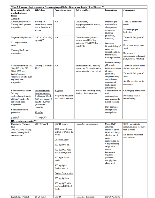
please click on the image for a larger view.
|
|
Patient Case #1
SG is a 68-year-old female who presents to a community pharmacy with complaints of heartburn and belching for the past several months. According to the patient, she takes a Tums when she thinks of it, but usually just has a glass of warm milk and lies down until it passes. Over the past week, even these interventions have not helped relieve symptoms. It is worse at night when she is lying on the recliner watching television. SG states that her friend told her to try OTC Prilosec.
- Is empiric therapy with a PPI indicated at this time?
- Yes, because she has a treatment failure to Tums
- Yes, because she has a presumptive diagnosis of GERD based on her symptoms
- No, because she has not implemented lifestyle modifications
- No, because she needs to have a documented diagnosis with endoscopy first
B) Endoscopy is not recommended for diagnosis of GERD, and pharmacologic therapy can be initiated as soon as the presumptive diagnosis of GERD is made. Lifestyle modifications should be initiated in all patients, and Tums is not considered an effective therapy for GERD.
- What lifestyle modifications should the pharmacist counsel the patient on?
- Avoidance of food triggers, such as mint, spicy foods, chocolate, and alcohol
- Patient should not recline, but should lie flat after eating
- The patient should consider losing 10 to 15 pounds
- Do not eat meals within 2 to 3 hours of bedtime
D) Recent data do not support reduction of GERD symptoms with food trigger avoidance, and therefore specific food avoidance is no longer recommended as part of lifestyle modifications. Patients should only lose weight if they recently gained weight and have a BMI >25 kg/m2. Patients should lie with their head slightly elevated, at least 2 to 3 hours after eating.
|
There have been concerns in recent years regarding the safety of PPIs in patients taking concomitant thienopyridines (e.g., Plavix) and those with osteoporosis.2,5 Similarly, concerns for increased risk of Clostridium difficile infection, and pneumonia have been newsworthy. However, according to the 2013 guidelines, PPI therapy may be continued in the presence of osteoporosis and concomitant clopidogrel, as the evidence does not support increased risk of hip fracture or cardiovascular events, respectively, unless additional risk factors exist. There is evidence that taking PPIs may increase the risk of C. difficile infection, and at-risk patients should be closely monitored or alternate therapies should be initiated. Furthermore, the risk of community-acquired pneumonia may be increased with short-term PPI therapy, though this risk is not evident in those who have been taking PPIs long-term.
|
Patient Case #1 continued
SG comes back to the pharmacy a year later after seeing a commercial highlighting the risk of osteoporosis with using drugs like Prilosec. Her past medical history includes GERD (diagnosed a year ago), osteoporosis (no previous fractures) diagnosed in 2012, and glaucoma diagnosed in 2009.
- How should her concerns about osteoporosis be addressed?
- Recent studies do not support any risk for osteoporosis with the long-term use of PPIs
- The use of long-term PPIs is appropriate in this patient
- The use of short-term PPIs increases the risk of osteoporosis
- The risk of osteoporosis is higher with H2RAs
B) This patient has osteoporosis; however, without additional risk factors for hip fracture, such as kidney disease, previous vertebral or hip fracture, diabetes, corticosteroid use, or osteoarthritis, long-term PPI use may be continued. Short-term PPI use may increase the risk of CAP.
|
Monitoring Parameters1,4
- Therapy-related adverse drug reactions should be monitored (Table 2)
- Management of drug interactions, particularly with antacids or cimetidine, should be proactively approached (Table 2)
- Renal/hepatic function should be monitored as some medications require dosage adjustment
- Assess effectiveness of therapy after 8 to 16 weeks
- Monitor for refractory and alarm symptoms (dysphagia, weight loss, GI bleeding, anemia) and refer patient to provider if they occur
PEPTIC ULCER DISEASE (PUD)
Overview
Peptic ulcers affect 4 million adults in the US each year, with a lifetime prevalence of 12% in men and 10% in women.6 Complications from peptic ulcer disease (PUD) result in approximately 15,000 deaths annually, and the estimated economic burden on the health system is around $6 billion per year. An estimated 70% of duodenal ulcers are due to the gram negative bacillus, Helicobacter pylori in Western countries.7 Regular nonsteroidal anti-inflammatory drug (NSAID) use increases the odds of gastrointestinal (GI) bleeding by 5- to 6-fold, resulting in serious complications in 1% to 4% of individuals using NSAIDs and 100,000 related hospital admissions annually.
Peptic ulcer disease encompasses ulcerations or erosions in the mucosal surface of the stomach and duodenum.6,7 There are 3 main types: H. pylori-induced, NSAID-induced, and stress-induced ulcers.8 Ulcers not caused by H. pylori or NSAIDs are uncommon, and are caused by gastric hypersecretion, other diseases, genetic predisposition, gastric outlet obstruction, or heavy tobacco use. Stress-induced ulcers in the critical care setting are beyond the scope of this review. Additional risk factors for PUD are in Table 3.
| Table 3. Risk Factors for Peptic Ulcer Disease5,6 |
- Medications
- Aspirin
- Bisphosphonates
- Chemotherapy
- Potassium chloride
- Mycophenolate mofetil
- Clopidogrel
- Smoking
- Emotional stress
|
The development of an ulcer occurs when there is an imbalance between increased exposure to gastric acid and pepsin and the protective mechanisms of the mucosa.8 Hydrochloric acid and pepsinogen are secreted from the parietal and chief cells, respectively. Pepsin is a cofactor that is involved in proteolysis and is activated optimally at a pH of 1.8 to 3.8, and irreversibly inactivated at a pH of 7.
The mucus and bicarbonate layers of the stomach have a nearly neutral pH and protect the stomach from acidic contents.8 Repair of the mucosal lining occurs with epithelial cell growth, and maintenance requires the production of prostaglandins via cyclooxygenase (COX)-1 and COX-2.7,8 H. pylori and NSAIDs affect the mucosal defenses through different mechanisms.7
H. pylori is a pH-sensitive bacterium that lives between the mucus layer and epithelial lining of the stomach or duodenum.8 H. pylori is able to create a microenvironment of protection from the acidic gastric contents. Gastric inflammation is produced via several processes, which ultimately leads to mucosal injury. It is transmitted by the oral-oral or oral-fecal routes and is always associated with active gastritis; however, only 10% to 15% of patients with H. pylori develop an ulcer.
Nonsteroidal anti-inflammatory drugs induce mucosal damage by directly irritating the epithelium and by inhibiting mucosal prostaglandin synthesis.8
Although pain often occurs after meals, presence or absence of such pain does not confirm or rule out an ulcer.8 Similarly, dyspepsia has not been shown to correlate with ulcers. Complications that are common with PUD are GI bleeding, perforation, and gastric outlet obstruction.
There are no routine laboratory tests that aid in the diagnosis of PUD.8 Diagnosis differs depending on the type of suspected ulcer. H. pylori infection is diagnosed with endoscopic or nonendoscopic testing to obtain 3 tissue samples from certain areas of the stomach.8,9 The biopsy is tested with a urease test, culture, polymerase chain reaction, or histology. Antibiotics and bismuth salts must be held for 4 weeks prior and PPIs held 1 to 2 weeks prior to biopsy, as these may affect the sensitivity of the rapid urease test used for diagnosis.8
Noninvasive testing such as the urea breath test, fecal antigen test, or serologic testing may be performed.6 Otherwise, documented ulcers are found via barium radiography or endoscopy. Endoscopy is used both for diagnosis and to rule out malignancy in gastric ulcers; duodenal ulcers are rarely malignant.10
Guidelines
The American Society for Gastrointestinal Endoscopy published guidelines for the role of endoscopy in PUD in 2010.10 The ACG published guidelines on the management of H. pylori infection in 2007 (an update is currently in progress).9 The goals of therapy are in Table 4.
| Table 4. Goals of Therapy for Peptic Ulcer Disease8 |
- Reduce pain
- Heal ulcer
- Prevent recurrence
- Minimize risk of complications from the ulcer
- Eradicate bacteria (Helicobacter pylori)
- Rapid healing (NSAID-induced)
|
| NSAID = nonsteroidal anti-inflammatory drug. |
Treatment
In addition to lifestyle modifications, pharmacologic therapy is necessary for the healing and prevention of ulcers (Table 2).4,8 The agents chosen are based on the cause of the ulcer.
Although an individual's diet is not likely to be the cause of a gastric ulcer, it is still recommended that certain foods be avoided due to their ability to exacerbate symptoms of ulcers.8 Examples of these foods are spicy foods, caffeine, and alcohol. Additionally, it is helpful to reduce the use of aspirin or NSAIDs, reduce or quit smoking, and lessen psychological stressors.
The 2007 ACG guidelines on the management of H. pylori infection recommends a clarithromycin-based triple therapy regimen for 14 days (although resistance to this regimen is on the rise) or a bismuth-based quadruple therapy regimen for 10 days (Table 5).9 The eradication rates are slightly higher in the US for the bismuth-based therapy at 75% to 90%, compared to the clarithromycin-based therapy at 70% to 85%. Antisecretory agents are necessary to both regimens since they increase gastric pH and decrease volume, which enhances the activity and concentration of the antibiotic.8 Additional considerations and full dosing details can be found in Table 2. A sequential regimen exists but has not been validated in the US, and therefore is not recommended as first-line therapy per the ACG guideline. In the case of treatment failure with a first-line regimen, an effort should be made to avoid previously trialed antibiotics.
| Table 5. H. pylori Regimens8,9 |
Triple Therapy
PPI once or twice daily
AND
Clarithromycin 500 mg BID
AND
Amoxicillin 1 g BID OR metronidazole 500 mg BID |
Quadruple Therapy
Bismuth subsalicylate 525 mg QID
AND
Metronidazole 250 mg or 500 mg QID
AND
Tetracycline 500 mg QID
AND
PPI OR H2RA once or twice daily |
| BID = twice daily; H2RA = histamine H2 receptor antagonist; QID = 4 times daily; PPI = proton pump inhibitor. |
The treatment of NSAID-induced ulcers is selected based on H. pylori status; therefore, H. pylori testing must first be performed.8 If there is no H. pylori detected, the NSAID should be discontinued and drug therapy with a PPI, H2RA, or sucralfate initiated. If the ulcer is positive for H. pylori, a 3-drug regimen including a PPI should be initiated for eradication.
Some patients cannot reasonably discontinue NSAID therapy, so these individuals should be managed with a PPI for potent acid suppression if H. pylori is absent, or with a PPI-based regimen if H. pylori is present.8 Some patients may require the addition of misoprostol, a prostaglandin analog, if they are at risk of developing a complication. Switching from an NSAID to a selective COX-2 inhibitor is another option for these patients.
Non-H. pylori, non-NSAID-induced ulcers are managed with either a PPI for 4 weeks or a H2RA or sucralfate for 6 to 8 weeks.8 Antacids alone are not recommended due to the high dose and duration of therapy required.
Monitoring Parameters8
For all recommended H. pylori regimens, it is important to monitor for adverse events and drug interactions, especially with the antibiotics used (Table 2). It is also essential to check for persistent symptoms of an ulcer at the end of the recommended treatment course. For patients with NSAID-induced ulcers, monitoring of upper GI complications as well as treatment-related adverse events and drug interactions should be performed. Resolution of ulcer pain should occur within a few days of discontinuation of NSAIDs and within a week of initiated therapy.
INFLAMMATORY BOWEL DISEASE (IBD)
Overview
Inflammatory bowel disease (IBD) consists of 2 conditions: 1) ulcerative colitis (UC), a mucosal inflammation of the rectum and colon, and 2) Crohn's disease (CD), a transmural inflammation affecting any part of the GI tract from mouth to anus.11
The highest rates of IBD are found in North America, Northern Europe, and Great Britain.11 Crohn's disease and UC have an incidence of 0.03 to 15.5 cases and 1.2 to 20 cases per 100,000 persons per year, respectively. The peak incidence for CD occurs earlier in life, in the 20th decade, whereas the peak incidence of UC occurs twice, in the 30s, and then in the 60s and 70s.
The etiology of IBD is unknown, but infectious, genetic, environmental, and immunologic factors are thought to be contributory (Table 6).11 The pathologic features differ between CD and UC (Table 7).11-13 Some patients experience extraintestinal complications including arthritis, ocular complications, skin and mucosal lesions, anemia, increased risk of venous thromboembolism, and bone disease.
| Table 6. Etiologic Factors of Inflammatory Bowel Disease11-13 |
|
Genetic factors → Predisposition to IBD
Microorganisms → Development of IBD
Immunologic factors → Pathogenesis of IBD
Psychological factors (stress) → Disease flares
Lifestyle → Smoking – protective for UC, worsens CD
NSAIDs – disease flares
|
| CD = Crohn's disease; IBD = inflammatory bowel disease; NSAID = nonsteroidal anti-inflammatory drug; UC = ulcerative colitis. |
| Table 7. Pathologic Features of Inflammatory Bowel Disease11-13 |
Ulcerative colitis
- Rectal involvement
- Crypt abscesses
- Superficial inflammation of mucosal and submucosal layers
- Friability
|
Crohn's disease
- Ileal involvement
- Strictures & fistulas
- Granulomas
- Linear clefts
- Transmural inflammation
- Cobblestone appearance
|
Symptoms of UC vary from abdominal cramping associated with frequent but small bowel movements to profuse, sometimes bloody, diarrhea.11 Frequency of bowel movements ranges from fewer than 4 stools in mild disease to greater than 6 in severe disease. Severe disease also manifests as bloody diarrhea with fever, leukocytosis, possible dehydration, tachycardia, and hypotension. For CD, symptoms also range in severity, and include diarrhea and abdominal pain, weight loss, and possible rectal bleeding.12 Perianal symptoms may occur before bowel symptoms do, which differentiates CD from UC. Fulminant disease is marked by toxic megacolon.
Diagnosis of CD is made with pathology, radiography, and endoscopy based on the presenting signs and symptoms.12 Other bowel diseases must be ruled out. Disease activity is defined and used for choosing the appropriate therapy. Definitions for remission, mild-moderate, moderate-severe, and severe-fulminant disease correspond to the Crohn's Disease Activity Index (CDAI).
Diagnosis of UC requires stool examinations, sigmoidoscopy or colonoscopy, and biopsy.13 Negative stool evaluation is needed to rule out infectious causes. Activity definitions guide therapy and are outlined by severity and location.
|
Patient Case #2
AG is a 29-year-old male with complaints of increased frequency of bowel movements (5 to 6 daily) for 2 weeks with some containing blood. He also has felt weaker over the past week. He reports having had similar bouts of diarrhea and weakness in the past couple of years, but none lasting this long. The patient reports being particularly stressed at work during this tax season. He has never traveled overseas and reports no recent sick contacts. He recently injured his lower back while moving and has been taking ibuprofen 3 to 4 times a day for the past 3 weeks, and denies taking any other medications, including antibiotics. He has been smoking 1 pack of cigarettes per day since his early 20s. The decision is made to perform lower GI endoscopy with biopsy.
The histologic findings include granulomas and strictures with evidence of transmural inflammation and cobblestone appearance, especially in the ileum. Crohn's disease is diagnosed.
- Based on AG's history of present illness, what counseling can you offer the patient at this time?
- Smoking cessation
- Appropriate use of NSAIDs
- Stress management
- All of the above
D) Smoking has been shown to worsen CD; NSAID use and stress are associated with disease flares. Along with pharmacologic therapies, the patient should be counseled on management of these triggers.
|
| Table 8. Goals of Therapy for Irritable Bowel Disease12,13 |
Ulcerative colitis
- Induce and maintain symptom remission
- Improve quality of life
- Reduce the need for chronic steroids
- Minimize cancer risk
|
Crohn's disease
- Eliminate disease-related symptoms
- Normalize quality of life
- Maintain overall well-being with few medication side effects and long-term complications
|
Guidelines
The American College of Gastroenterology published the Management of Crohn's Disease in Adults in 2009 and Ulcerative Colitis Practice Guidelines in 2010.12,13 Both of these guidelines are currently in the process of being updated.
Treatment
Patients with IBD require nutritional interventions to help manage the malnourishment that often occurs in these patients.11 Malabsorption also may result from small bowel involvement of patients with CD, since many nutrients are absorbed at this site. Diets intended to help minimize symptoms are not typically recommended, unless the individual also has lactose intolerance, which is common in patients with IBD. Enteral feeding may also be used for patients in acute or chronic situations.
According to the 2 guidelines for CD and UC, the location and severity of disease determines the pharmacological therapy (including dose, route, frequency, and formulation) (Table 9).4,12-14 Several classes of drugs are recommended, including aminosalicylates, corticosteroids, immunosuppressive agents, antimicrobials, tumor necrosis factor (TNF)-alpha inhibitors, and an integrin receptor adhesion inhibitor.12-15 Although not discussed in the current CD and UC guidelines, vedolizumab (Entyvio) was approved by the Food and Drug Administration (FDA) in May 2014 to treat adults with moderate to severe UC and CD when 1 or more standard therapies (corticosteroids, immunomodulators, or TNF-alpha inhibitors) have not resulted in an adequate response.16
|
Table 9. Guideline-Recommended Drugs for Ulcerative Colitis and Crohn's Disease4,11-14*
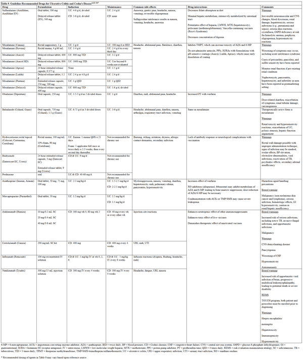
please click on the image for a larger view.
|
Ulcerative Colitis
The same drugs are used for induction of mild to moderate distal and extensive disease.13 Both oral and topical routes of administration are effective, and thus the patient's preference plays a role in the selection of agent. Topical mesalamine is considered first-line and is superior to oral aminosalicylates alone. Some patients require combination topical and oral therapy for maximal effect. If aminosalicylates do not succeed in inducing remission, alternative treatment options include oral corticosteroids, infliximab, 6-mercaptopurine, or azathioprine.
Immunomodulators and TNF-alpha inhibitors have a first-line role in the induction of severe disease. Maintenance regimens include the aminosalicylates for distal and extensive disease, with azathioprine or 6-mercaptopurine as second-line agents. Corticosteroids should not be used chronically. Infliximab may also be used for maintenance of distal and extensive disease.
Crohn's Disease
Sulfasalazine or budesonide, if confined to the ileum or right colon, is recommended for induction of mild to moderate Crohn's disease.12 However, these agents do not consistently work for maintenance therapy. Mesalamine was often used in the past, though recent evidence suggests it is not more effective than placebo for remission induction. Moderate to severe disease may require initial therapy with corticosteroids. Azathioprine/6-mercaptopurine and methotrexate have demonstrable benefits after inductive therapy with corticosteroids for maintenance of remission after steroid use. Tumor necrosis factor-alpha inhibitors are options if the first-line agents do not induce a response, or if patients have a contraindication to corticosteroids. Maintenance of remission may be managed with these agents, as well as with natalizumab. Severe, fulminant disease requires hospitalization, parenteral medications, surgical evaluation, and nutritional support. Perianal disease may require antibiotics, immunosuppressants, infliximab, and surgical drainage.
Traditionally, a "bottom-up" approach to treatment has been used, in which symptoms are managed first with aminosalicylates and corticosteroids, before stepping up to immunosuppressant and biologic agents if response is poor.11 There has been recent interest in the opposite approach due to the potential for altering the course of the disease; however, this method would be costly and may effectively eliminate any alternative therapy in the setting of early treatment failure.
|
Patient Case #2 continued
2. The patient is diagnosed with ileal mild to moderate active Crohn's disease. What is an appropriate first-line therapy for AG?
- Infliximab 5 mg/kg IV on weeks 0, 2, 6
- Mesalamine rectal suppository once daily
- Mesalamine 24-hour extended release capsule (Apriso) 1.5 g by mouth daily
- Budesonide extended release capsule 9 mg daily
D) Mesalamine is no longer recommended for use in CD. It is a first-line therapy for patients with UC. The patient is diagnosed with ileal mild to moderate active CD. Infliximab is not recommended for mild disease.
3. After AG responded to initial therapy, which is the best maintenance regimen?
- Continue budesonide therapy
- Switch to prednisone 40 mg by mouth once daily
- Azathioprine 2 mg/kg daily
- Sulfasalazine 3 g daily in divided doses
C) Long-term corticosteroid use beyond 6 months is not recommended due to the potential for side effects. Sulfasalazine is not used for maintenance therapy of CD.
4. What counseling is necessary for the regimen chosen?
The patient should be counseled on the potential for myelosuppression, rash, non-melanoma skin cancer and lymphoma, and risk of serious infections such as tuberculosis or invasive fungal infections.
|
Monitoring Parameters
- Adverse effects, especially with immunosuppressants, corticosteroids, and sulfasalazine (Table 9)
- Hematologic labs and monitoring for certain infections with use of immunosuppressants and TNF-alpha inhibitors
VIRAL HEPATITIS
Overview
There are 5 known types of hepatitis viruses; this review will focus on hepatitis B (HBV) and hepatitis C (HCV).17 Viral hepatitis can be both acute or chronic, though hepatitis A and E are self-limiting and do not cause chronic hepatitis.18 The epidemiology and etiology of hepatitis A virus, HBV, and HCV are in Table 10.
| Table 10. Epidemiology and Etiology of Viral Hepatitis17-19 |
| |
Hepatitis A |
Hepatitis B |
Hepatitis C |
| Type |
Acute |
Acute and chronic |
Acute and chronic |
| Reported/ Estimated New Cases (2013) |
1,781/3,500 |
3,050/19,800 |
2,138/29,700 |
| Transmission |
Oral-fecal |
Sexual, percutaneous exposure to blood, perinatal, close person-person contact |
Percutaneous exposure to blood |
| Risk factors |
- International travel
- Children
- Sexual/household contact with infected person
- MSM
- IDU
- Chronic liver disease
|
- Multiple sexual partners
- MSM
- IDU
- Sexual/household contact with infected person
- Co-infected with HIV or HCV
- Inmates
- Patients on hemodialysis
|
- IDUa
- Homeless
- Prisoners
- Intranasal cocaine
- Multiple sexual partners and coinfection with STD
- Receipt of blood products pre-1992 or clotting factors pre-1987
- Long-term hemodialysis
- Needle-stick exposure (healthcare workers)
|
| Incubation time |
28 days |
6 weeks to 6 months |
4 weeks to 12 weeks |
HCV = hepatitis C virus; HIV = human immunodeficiency virus; IDU = intravenous drug user; MSM = men who have sex with men; STD = sexually transmitted disease.
a Most important risk factor |
Initially, the hepatocytes are infected and an incubation period of rapid viral replication occurs.19 Antigens and antibodies to the virus accumulate in urine, stool, and body fluids. Most patients are asymptomatic during this phase, but nonspecific symptoms such as anorexia, weakness, nausea/vomiting, jaundice, dark urine, and pale stools may occur.18 Hepatocyte death prompts an inflammatory response that leads to laboratory changes and symptoms of liver disease.19
Persistence of hepatitis viruses may lead to an immune-mediated chronic disorder of the liver.19 Histopathologic findings of chronic hepatitis show mononuclear cell infiltration into the portal areas as well as hepatocyte necrosis in the adjacent parenchyma, which can progress to fibrosis. Persistent inflammation and damage from any cause, fibrosis, and attempts at regeneration of the liver defines cirrhosis. As the liver tissue loses function, the liver decompensates and results in encephalopathy, development of varices, and decreased bile flow.
Chronic HBV is diagnosed when the hepatitis B surface antigen (HBsAg) has been positive for more than 6 months and the serum HBV DNA exceeds 20,000 IU/mL.20 Liver enzymes are often elevated and liver biopsy shows inflammation. Once antibodies to HBsAg have developed, the individual is considered to have immunity to HBV.18 Hepatitis B e antigen (HBeAg) is present in acute infections, marks viral replication, and is used to determine the appropriate regimen. Inactive HBV carriers and those with resolved HBV infection have specific diagnostic criteria as well, outlined in the guidelines.20 Unlike HCV, genotyping is not recommended as standard practice at this time.
Presence of HCV antibody indicates an active, acute, or chronic infection, past resolved infection, or false-positive result.21 A HCV nucleic acid test must be used to confirm the infection and guide therapy. In addition to obtaining a viral load (HCV RNA), HCV genotyping must be completed prior to initiating therapy.
Guidelines
The American Association for the Study of Liver Diseases (AASLD) published an updated guideline for the treatment of chronic HBV in 2015.21 The AASLD in conjunction with the Infectious Diseases Society of America (IDSA) is constantly updating the Recommendations for Testing, Managing, and Treating Hepatitis C as new information becomes available.22 This living document may be found at http://www.hcvguidelines.org/ and pharmacists and APRNs should regularly visit this site for the most current information regarding HCV management.
Treatment
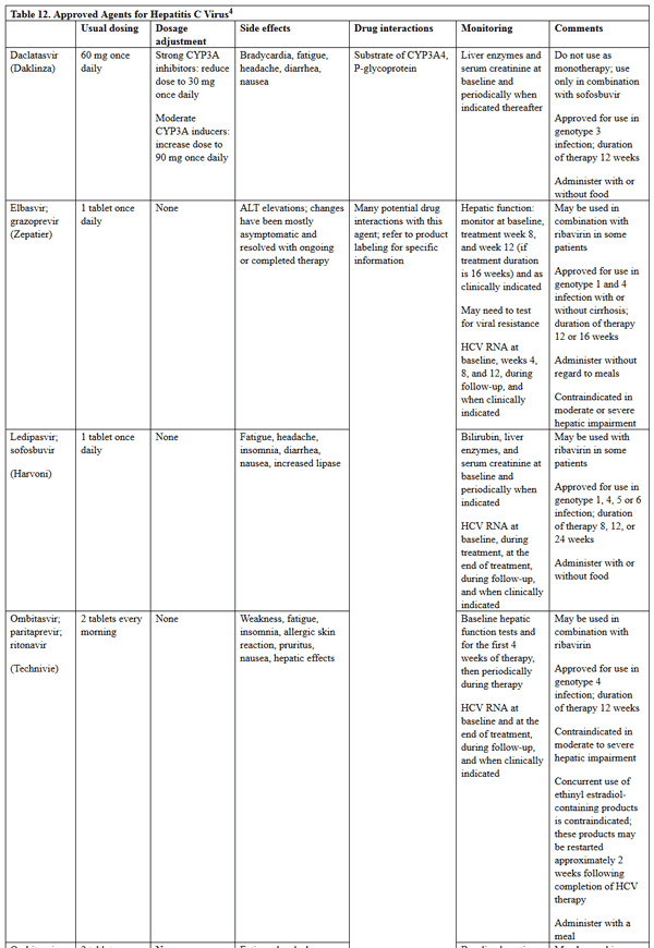
Pharmacologic therapy differs between HBV and HCV, and specific drug information for the agents used is in Tables 11 and 12.4,21 Of note, the treatment of HCV infection has undergone a major paradigm shift since 2011 when pegylated interferon and ribavirin was the standard of care for most patients. Table 12 includes information on only the direct-acting antivirals that have more recently become first line therapy for patients with HCV infection; no information on ribavirin or interferon products is included. Additionally, both telaprevir and boceprevir have either been removed from the US market or will be removed from the market in the near future; therefore, information on these products was also excluded.
|
Table 11. Approved Agents for Hepatitis B Virus4,21
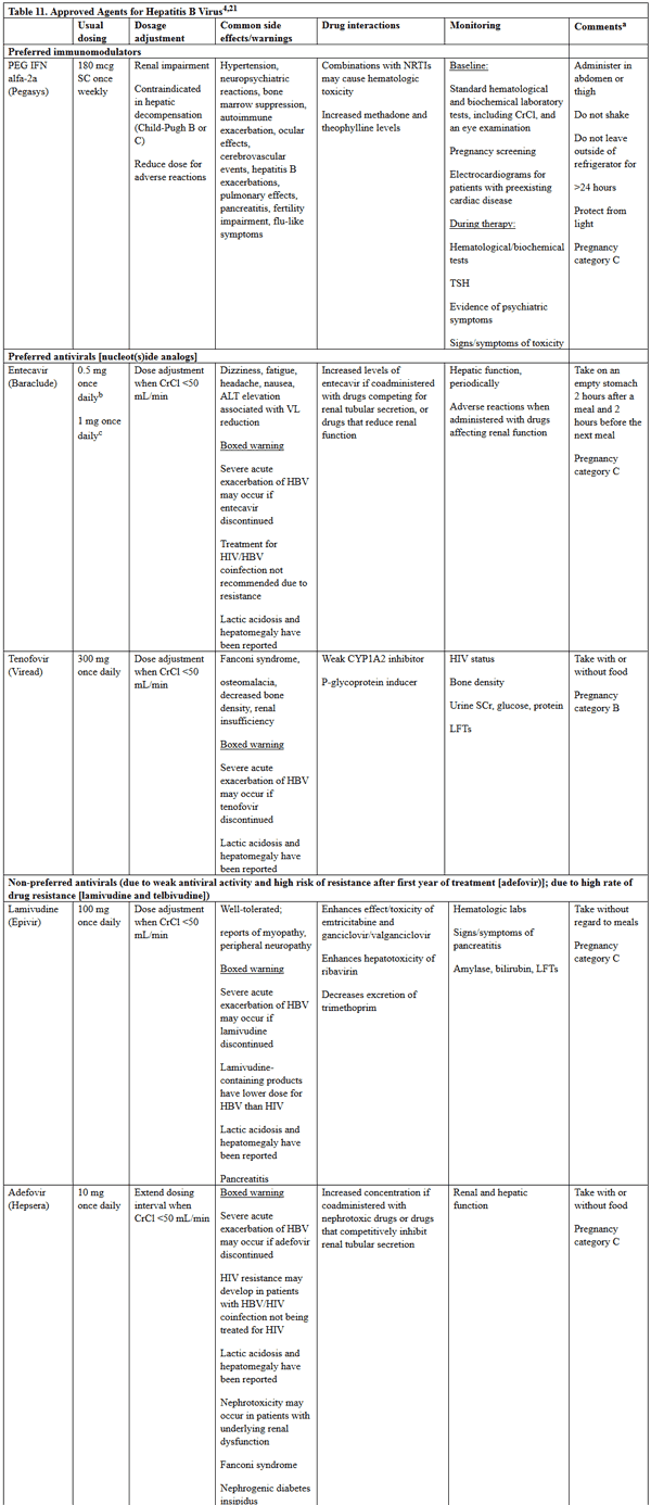
please click on the image for a larger view.
|
|
Table 12. Approved Agents for Hepatitis C Virus4
please click on the image for a larger view.
|
Hepatitis B Virus
Hepatitis B virus is not curable, and goals of therapy are in Table 13.21 Although there are 7 Food and Drug Administration (FDA)-approved antiviral medications for HBV, tenofovir and entecavir are the preferred nucleos(t)ide analogs due to increased resistance to the other options. Pegylated interferon (PEG-IFN) is preferred to regular interferon as it is more convenient to dose and has fewer adverse effects. Therapy is monitored by treatment response as defined in Table 14. Details on specific treatment regimens may be found in the most recent AASLD guidelines for treatment of chronic hepatitis B, which can be freely accessed at: https://www.aasld.org/sites/default/files/guideline_documents/hep28156.pdf.
| Table 13. Goals of Therapy for Hepatitis B Virus Infection21 |
- Achieve sustained suppression of HBV replication and remission of liver disease
- Prevent long-term complications, such as cirrhosis, hepatic failure, and hepatocellular carcinoma
|
| HBV = hepatitis B virus |
| Table 14. Definitions of Response to Treatment of Hepatitis B Virus Infection20 |
| Biochemical |
Decreased ALT to normal range |
| Histologic |
Decrease in histology activity index by at least 2, and no worsening of fibrosis |
| Virologic |
Decreased HBV DNA to undetectable, and a loss of HBeAg if initially positive |
| ALT = alanine aminotransferase; HBeAg = hepatitis B e antigen; HBV = hepatitis B virus. |
Hepatitis C Virus
According to the AASLD/IDSA guideline, treatment is recommended for all patients with chronic HCV infection, except those with short life expectancies that cannot be remediated by treating HCV, by transplantation, or by other directed therapy.22 Details on specific HCV treatment regimens are rapidly changing; therefore, pharmacists and APRNs should consistently refer to http://www.hcvguidelines.org/ when questions regarding appropriate treatment options arise.
Monitoring
- Drug-related adverse effects and interactions, including drug-food and drug-disease state interactions (Tables 11 and 12)
- Response to therapy as a marker of disease progression
Focus Points for Medication Therapy Management (MTM) in GI Disorders
GERD
Patient counseling
- Ensure patient is using over-the-counter (OTC) dosing unless they have been evaluated for GERD by a health care practitioner (Table 2).
- Counsel patient on appropriate timing of drug administration (Table 2), lifestyle modifications, intended goals and duration of therapy, and when to consult a physician for alarm symptoms.
- Monitor adherence to therapy and adverse events, and screen for potential drug-drug interactions.
PUD
Patient counseling
- Due to the possibility of treatment resistance to the H. pylori regimens, it is of utmost importance to counsel patients to adhere to the full regimen.
- In patients with NSAID-induced ulcers, explanation of risk and GI complications should be given.
- Explanation of why each medication is used may be helpful to include in addition to expected adverse events and timing of medication administration (Table 2).
- If alarm symptoms occur (bloody or black tarry stools, vomiting, severe abdominal pain, returning symptoms after H. pylori eradication), the patient should contact their provider for evaluation.
- Female patients of childbearing age should be counseled on possible reduced efficacy of oral contraceptives with the use of antibiotics.
IBD
Patient counseling
- Appropriate drug use and assess patient understanding of use
- Aminosalicylate agents have slow onset of action (2 to 4 weeks for effect)
- Azathioprine and 6-mercaptopurine can take 3 to 6 months to have optimal effect
- May refer patients to Crohn's and Colitis Foundation of American (CCFA) at www.ccfa.org
- Emphasize the importance of adherence to medications and follow-up visits, which helps reduce need for other costly medical services
Health maintenance
- Bone health
- Due to corticosteroid use or malabsorption
- High-risk patients should be screened for osteoporosis (i.e., previous vertebral fracture, postmenopausal females, males >50 years of age, chronic corticosteroid users, hypogonadism)
- Calcium/vitamin D supplementation or bisphosphonate may be appropriate
- Nutritional
- Iron, folate, vitamin B12 deficiency
- Sulfasalazine users should have daily folate supplementation
- Supplement B12 in patients with CD in the small bowel (ileocolic or gastric)
- Iron deficiency due to blood loss and inflammation
- Consider dietitian consultation
- Colorectal cancer screening
- For UC, annual or biannual colonoscopy is recommended in patients with 8 to 10 years of colitis. For CD, the guideline is silent on the issue of colorectal cancer screening.
- Vaccinations
- Since patients are often on immunosuppressive therapy, immunization against hepatitis A and B, influenza, tetanus, pneumonia, diphtheria, pertussis, and varicella is recommended
- These vaccines should be given prior to initiation of immunomodulator therapy
- Pregnancy and fertility
- Immunomodulators can decrease fertility in men
- Clinical improvement occurs during pregnancy, but increased risk of preterm birth in CD specifically
- Immunomodulator use controversial
Viral Hepatitis
Counseling for HBV transmission prevention
- Do not share toothbrushes or razors.
- Clean blood spills with bleach.
- Cover open wounds.
- Do not donate organs, sperm, or blood.
- Sexual/household contacts should receive HBV vaccination.
- Use barrier protection if sexual partner is not vaccinated or naturally immune.
- At-risk patients should be tested for appropriate response to vaccine series.
- Abstinence or limited alcohol use in HBV carriers, as heavy use (>20 g/d in women and >30 g/d) in men may increase risk of development of cirrhosis.
- Pregnant women who are HBsAg positive should inform their physician so that appropriate measures can be taken to prevent perinatal transmission (e.g., use of immune globulin and HBV vaccine in the newborn).
Counseling for prevention of HCV transmission and reduction of liver disease progression
- Do not share toothbrushes, dental, or shaving materials and cover any bleeding wounds.
- Cessation of intravenous drug use, or avoid reusing/sharing syringes, cotton, needles.
- Use of barrier protection for men who have sex with men coinfected with HIV who have multiple sexual partners.
- Do not donate blood, and clean any visible blood spills from an HCV infected person with 1:9 parts bleach to water.
- Abstinence from alcohol, as >50 g of alcohol daily increases risk of worsening fibrosis.
- Evaluation for coinfection with HBV or HIV, as these are associated with worse prognosis of HCV.
- Evaluate for advanced fibrosis or hepatocellular carcinoma (HCC).
- HCV testing
- Recommended if born between 1945 and 1965, and high risk behaviors, risk exposures, or unexplained liver disease or HIV infection.
Methods for overcoming barriers to HCV treatment
- Utilize referral services for patients with contraindications to treatment.
- Engage case managers, primary care, or medical homes to help make treatment and follow-up a priority for patients.
- Optimize regimens with better tolerability or simple instructions to help patients manage the long treatment duration or adverse events that occur with therapy.
- Create models of care that involve collaboration with pharmacists, specialists, and primary care providers to aid in accessibility to treatment.
- Utilize patient assistance programs for patients struggling with the high cost of therapy.
- Develop performance measures and accessible HCV guidelines to help improve provider expertise on HCV therapy.
References
- May DB, Rao S. Gastroesophageal reflux disease. In: DiPiro JT, Talbert RL, Yee GC, et al, eds. Pharmacotherapy: A Pathophysiologic Approach. 9th ed. New York, NY: McGraw-Hill; 2014. http://accesspharmacy.mhmedical.com.proxy.cc.uic.edu/content.aspx?bookid=689&Sectionid=45310475. Accessed April 25, 2016.
- Katz PO, Gerson LB, Vela MF. Guidelines for the diagnosis and management of gastroesophageal reflux disease. Am J Gastroenterol. 2013;108(3):308-328.
- Anon. Drugs for peptic ulcer disease and GERD. Treat Guidel Med Lett. 2014;12(140):25-30.
- Wickersham RM, ed. Drug Facts and Comparisons. St. Louis, MO: Wolters Kluwer Health; 2016. http://online.factsandcomparisons.com/index.aspx. Accessed April 27, 2016.
- Abraham NS, Hlatky MA, Antman EM, et al; ACCF/ACG/AHA. ACCF/ACG/AHA 2010 expert consensus document on the concomitant use of proton pump inhibitors and thienopyridines: a focused update of the ACCF/ACG/AHA 2008 expert consensus document on reducing the gastrointestinal risks of antiplatelet therapy and NSAID use. A Report of the American College of Cardiology Foundation Task Force on Expert Consensus Documents. J Am Coll Cardiol. 2010;56(24):2051-2066.
- Del Valle J. Peptic ulcer disease and related disorders. In: Longo DL, Fauci AS, Kasper DL, et al, eds. Harrison's Principles of Internal Medicine. 19th ed. New York, NY: McGraw-Hill; 2015. http://accesspharmacy.mhmedical.com/content.aspx?sectionid=79747602&bookid=1130&Resultclick=2. Accessed April 26, 2016.
- Chan FKL, Lau JYW.. Peptic ulcer disease. In: Feldman M, Friedman LS, Brandt LJ, eds. Sleisenger and Fordtran's Gastrointestinal and Liver Disease: Pathophysiology, Diagnosis, Management. 10th ed. Philadelphia, PA: Saunders; 2016. https://www.clinicalkey.com/#!/content/book/3-s2.0-B9781455746927000533. Accessed April 26, 2016.
- Love B, Thoma MN. Peptic ulcer disease. In: DiPiro JT, Talbert RL, Yee GC, et al, eds. Pharmacotherapy: A Pathophysiologic Approach. 9th ed. New York, NY: McGraw-Hill; 2014. http://accesspharmacy.mhmedical.com.proxy.cc.uic.edu/content.aspx?bookid=689&Sectionid=48811467. Accessed April 26, 2016.
- Chey WD, Wong BC; Practice Parameters Committee of the American College of Gastroenterology. American College of Gastroenterology guideline on the management of Helicobacter pylori infection. Am J Gastroenterol. 2007;102(8):1808-1825.
- Banerjee S, Cash BD, Dominitz JA, et al; for the ASGE Standards of Practice Committee. The role of endoscopy in the management of patients with peptic ulcer disease. Gastrointest Endosc. 2010;71(4):663-668.
- Hemstreet BA. Inflammatory bowel disease. In: DiPiro JT, Talbert RL, Yee GC, et al, eds. Pharmacotherapy: A Pathophysiologic Approach. 9th ed. New York, NY: McGraw-Hill; 2014. http://accesspharmacy.mhmedical.com/content.aspx?bookid=689&Sectionid=45310476. Accessed April 26, 2016.
- Lichtenstein GR, Hanauer SB, Sandborn WJ; Practice Parameters Committee of American College of Gastroenterology. Management of Crohn's disease in adults. Am J Gastroenterol. 2009;104(2):465-483.
- Kornbluth A, Sachar DB; Practice Parameters Committee of the American College of Gastroenterology. Ulcerative colitis practice guidelines in adults: American College Of Gastroenterology, Practice Parameters Committee. Am J Gastroenterol. 2010;105(3):501-523.
- Anon. Drugs for inflammatory bowel disease. Treat Guidel Med Lett. 2012;10(115):19-28.
- Terdiman JP, Gruss CB, Heidelbaugh JJ, et al; AGA Institute Clinical Practice and Quality Management Committee. American Gastroenterological Association Institute guideline on the use of thiopurines, methotrexate, and anti-TNF-α biologic drugs for the induction and maintenance of remission in inflammatory Crohn's disease. Gastroenterology. 2013;145(6):1459-1463.
- Entyvio [package insert]. Deerfield, IL: Takeda Pharmaceuticals America, Inc.; 2014.
- Viral hepatitis. Centers for Disease Control and Prevention. www.cdc.gov/hepatitis. Updated May 31, 2015. Accessed April 26, 2016.
- Deming P. Viral hepatitis. In: DiPiro JT, Talbert RL, Yee GC, Matzke GR, et al, eds. Pharmacotherapy: A Pathophysiologic Approach. 9th ed. New York, NY: McGraw-Hill; 2014. http://accesspharmacy.mhmedical.com.proxy.cc.uic.edu/content.aspx?bookid=689&Sectionid=48811468. Accessed April 26, 2016.
- Khalili M, Burman B. Liver disease. In: McPhee SJ, Hammer GD. eds. Pathophysiology of Disease. 7th ed. New York, NY: McGraw-Hill; 2014. http://accesspharmacy.mhmedical.com/content.aspx?sectionid=53555695&bookid=961&jumpsectionID=53631433&Resultclick=2. Accessed April 26, 2016.
- Lok ASK, McMahon BJ. Chronic hepatitis B: update 2009. https://www.aasld.org/sites/default/files/guideline_documents/ChronicHepatitisB2009.pdf. Accessed April 26, 2016.
- Terrault NA, Bzowej NH, Change KM, Hwang JP, Jonas MM, Murad MH. AASLD guidelines for treatment of chronic hepatitis B. https://www.aasld.org/sites/default/files/guideline_documents/hep28156.pdf. Accessed April 26, 2016.
- AASLD and IDSA. Recommendations for Testing, managing, and treating hepatitis C. http://www.hcvguidelines.org/. Updated February 24, 2016. Accessed April 26, 2016.
Back to Top